Nuclear medicine uses small amounts of radiotracers to evaluate how organs and tissues function at the molecular level. Unlike conventional imaging, it provides detailed information about metabolic activity, helping physicians assess how cancer responds to treatment. This functional imaging supports treatment planning by identifying changes that may not be visible on standard imaging studies. Here is more information on nuclear medicine and its use in cancer therapy monitoring:
Imaging Function
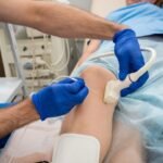
Nuclear medicine imaging uses small amounts of radioactive material called radiotracers to evaluate organ and tissue function. A healthcare provider administers the radiotracer by injection or, in some cases, orally, depending on the procedure. The radiotracer travels through the bloodstream and collects in targeted areas to support detailed imaging.
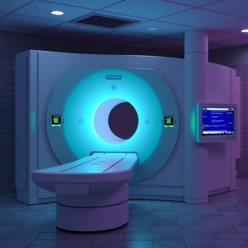
Specialized imaging equipment detects the radiotracer and creates images that show biological and metabolic activity within the body. This approach provides functional information that complements traditional imaging focused on anatomy. The additional information helps physicians evaluate disease processes and guide treatment planning.
PET Detection
Positron emission tomography (PET) is a nuclear medicine imaging technique used to evaluate organ and tissue function. PET scans help physicians identify abnormal cellular activity and are commonly used during cancer diagnosis, staging, and treatment monitoring. The information gathered complements findings from other imaging studies.
PET imaging works by detecting areas where radiotracers collect within the body. Many cancers show increased metabolic activity, allowing physicians to identify areas that may require further evaluation. Follow-up PET scans also help assess how tissues respond during the course of treatment.
PET scans are often combined with CT or MRI during the same appointment. These combined studies provide both functional and anatomical information in a single examination. The additional detail helps physicians develop more informed diagnostic and treatment plans.
Bone Scans
Bone scans are nuclear medicine imaging studies that help physicians evaluate bone health during cancer care. They are commonly used to determine whether certain cancers have spread to the skeletal system and to investigate areas of abnormal bone activity. Bone scans may help evaluate:
- The spread of cancer to the bones (bone metastasis)
- Areas of abnormal bone activity
- Bone changes related to cancer treatment
- Fractures that may not appear on other imaging studies
- Bone infections or inflammation
- Arthritic changes affecting the bones and joints
The patterns seen on a bone scan help physicians distinguish between different causes of abnormal bone activity. This information supports treatment planning and helps determine whether symptoms are related to cancer, treatment effects, or another underlying condition.
Thyroid Imaging
Thyroid uptake scans evaluate thyroid function and detect conditions such as hyperthyroidism, hypothyroidism, inflammation, and nodules. Physicians also use these scans during cancer treatment because some therapies can affect thyroid activity over time. Providers administer a small amount of radiotracer to measure how much the thyroid absorbs and assess its level of function. Early detection of changes helps care teams adjust treatment and support ongoing patient health.
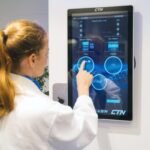
Schedule With Nuclear Medicine Today
Nuclear medicine provides detailed imaging that helps physicians understand how the body responds to cancer treatment. Techniques such as PET scans and bone scans allow providers to evaluate metabolic activity and detect the spread of disease. Thyroid and other nuclear imaging studies also help monitor organ function during ongoing therapy. Schedule a consultation with a nuclear medicine specialist today to determine which imaging options are appropriate for your care plan.